English
 Arabic
 French
 Spanish
 Portuguese
 Deutsch
 Turkish
 Dutch
Dutch
 Italiano
 Russian
 Romaian
 Portuguese (Brazil)
 Greek
Greek
Global Expandable Interbody Fusion System Market Forecast and Competitive Analysis 2034
Health |
2026-08-05 12:30:46
 Advertise With Us
 About Us
 Terms And Conditions
 Politique de confidentialité
Tout voir
Politique de confidentialité
Tout voir

Mise à niveau vers Pro
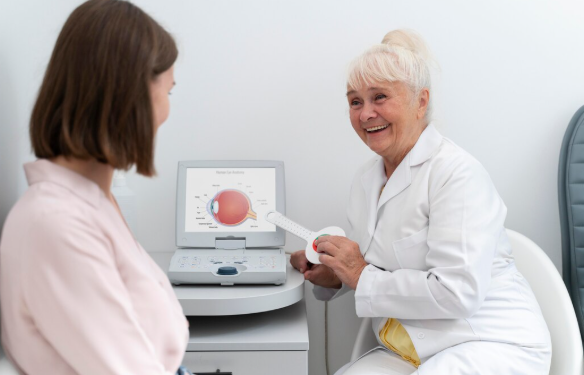
Imagine you've scheduled a breast health appointment and hear two terms: Mammogram Screening and breast ultrasound. They may sound similar, but they serve different purposes. Many women assume they are interchangeable, while others believe having one test means they no longer need the other. In reality, each imaging method provides unique information, and understanding their differences can help you make informed decisions about your breast health.
Knowing when Mammogram Screening is used, how breast ultrasound works, and why healthcare providers sometimes recommend both can reduce confusion and improve confidence during the screening process.
Breast imaging allows healthcare professionals to examine tissue that cannot be evaluated through visual inspection or physical examination alone. Many breast changes develop silently, making imaging an important part of preventive healthcare.
The goals of breast imaging include:
Detecting abnormalities before symptoms appear
Evaluating breast lumps or unusual changes
Monitoring previously identified findings
Comparing images over time
Supporting early diagnosis when additional evaluation is needed
Different imaging techniques are selected based on the individual's age, symptoms, breast density, and medical history.
Mammogram Screening is a preventive X-ray examination performed on women who usually do not have noticeable breast symptoms. It is designed to identify very small changes within breast tissue before they become visible or can be felt.
The examination uses low-dose X-rays to produce detailed images that may reveal:
Tiny calcifications
Small masses
Tissue distortion
Early structural changes
Areas requiring closer evaluation
Routine Mammogram Screening is considered one of the primary tools for early breast cancer detection.
A breast ultrasound uses sound waves rather than radiation to create images of breast tissue.
During the examination:
A clear gel is applied to the skin.
A handheld device called a transducer moves across the breast.
Sound waves produce images on a monitor.
The procedure is painless and does not involve compression like a mammogram.
Ultrasound is commonly used to provide additional information after a mammogram or to evaluate a specific breast concern.
Although both examine breast tissue, Mammogram Screening and breast ultrasound have different purposes.
Mammogram Screening
Uses low-dose X-rays
Produces detailed internal breast images
Excellent for detecting tiny calcifications
Breast Ultrasound
Uses sound waves
No radiation exposure
Excellent for evaluating fluid-filled and solid structures
Mammogram Screening
Routine preventive screening
Early detection before symptoms appear
Breast Ultrasound
Further evaluation of specific findings
Assessment of breast lumps or abnormalities
Additional imaging for dense breast tissue when appropriate
Mammogram Screening often identifies:
Small calcifications
Early tissue changes
Tiny abnormalities not easily felt
Breast ultrasound is particularly helpful for:
Distinguishing cysts from solid masses
Evaluating localized breast concerns
Examining certain areas more closely
Many women wonder why additional ultrasound is suggested after a Mammogram Screening.
The reason is simple: the two tests provide different types of information.
For example:
A mammogram may identify an area requiring closer examination.
Ultrasound can provide additional detail about that specific area.
Together, the tests offer a more complete evaluation.
Having both tests does not necessarily indicate a serious problem. Often, ultrasound is simply used to clarify what was seen on the mammogram.
This is one of the most common questions.
The answer depends on the reason for imaging.
Routine preventive screening
Detecting very early breast changes
Monitoring breast health over time
Comparing previous mammograms
Evaluating a newly discovered lump
Examining dense breast tissue in certain situations
Investigating localized pain or swelling
Providing additional information after mammography
Rather than competing, the two tests often complement each other.
Understanding the process can reduce anxiety.
The appointment generally includes:
Basic health information is reviewed.
You change into a gown from the waist up.
Each breast is positioned individually.
Gentle compression improves image quality and reduces movement.
Several images are taken from different angles.
The appointment usually lasts about 20 to 30 minutes.
The ultrasound experience differs from a mammogram.
Typical steps include:
Lying comfortably on an examination table
Application of warm gel
Moving the transducer over the breast
Viewing images on a monitor
Completing the examination within approximately 15 to 30 minutes
There is no breast compression during ultrasound.
Routine Mammogram Screening offers several important benefits.
These include:
Early detection of breast abnormalities
Ability to identify microscopic calcifications
Long-term comparison of breast images
Widely used preventive screening method
Quick examination
Minimal recovery time
Regular screening plays an important role in preventive breast healthcare.
Ultrasound also offers valuable benefits.
These include:
No radiation exposure
Comfortable examination
Helpful evaluation of breast lumps
Additional assessment after mammography
Useful for examining certain types of breast tissue
Real-time imaging
Each imaging method contributes valuable information in different situations.
Several myths create confusion.
Ultrasound is generally considered a complementary test rather than a replacement for routine mammography.
Although highly effective, mammograms cannot detect every abnormality, particularly in some women with dense breast tissue.
Ultrasound relies on sound waves, not radiation.
Being asked to undergo ultrasound after Mammogram Screening does not automatically indicate cancer.
Many follow-up examinations identify harmless findings.
Preparation for breast imaging is usually straightforward.
Helpful tips include:
Avoid deodorant and powder.
Wear a two-piece outfit.
Bring previous mammogram records.
Inform staff about breast implants or surgery.
Wear comfortable clothing.
Bring prior imaging reports if available.
Inform the technologist about current symptoms.
Good preparation helps ensure efficient imaging and accurate interpretation.
Imaging is only one component of preventive breast care.
Healthy lifestyle habits remain important throughout adulthood.
Consider:
Maintaining a healthy weight
Exercising regularly
Eating a balanced diet
Avoiding tobacco
Limiting alcohol
Managing stress
Getting sufficient sleep
Reporting breast changes promptly
Attending routine medical appointments
These habits work together with Mammogram Screening to support overall breast wellness.
Understanding the differences between Mammogram Screening and breast ultrasound helps eliminate confusion about breast imaging. While Mammogram Screening remains one of the primary methods for detecting early breast changes before symptoms appear, ultrasound provides valuable additional information when evaluating specific concerns. Rather than replacing one another, these imaging tests often work together to provide a more complete assessment of breast health.
By staying informed, following recommended screening schedules, and discussing individual risk factors with your healthcare provider, you can make confident decisions that support long-term breast health.
Yes. Depending on the reason for imaging and scheduling availability, both tests may sometimes be performed during the same visit.
Very little preparation is needed. Wearing comfortable clothing and bringing previous imaging records is usually sufficient.
The timing varies depending on the imaging facility. A radiologist typically reviews the images before the final report is provided.
Yes. A normal ultrasound does not replace routine Mammogram Screening, as both tests evaluate breast tissue differently and serve different purposes.