English
 Arabic
 French
 Spanish
 Portuguese
 Deutsch
 Turkish
 Dutch
Dutch
 Italiano
 Russian
 Romaian
 Portuguese (Brazil)
 Greek
Greek
Can Hisense TV Service Center repair damaged TV panels and screen problems?
Other |
2026-07-01 06:37:19
 Advertise With Us
 About Us
 Terms And Conditions
 Política de privacidade
Ver todos
Política de privacidade
Ver todos

Atualizar para Plus
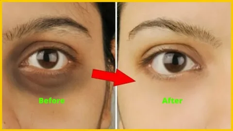
Dark circle treatment in Jeddah involving the correction of tear trough hollows with dermal fillers demands an absolute mastery of periorbital micro-anatomy and precision injection geometries. The tear trough, or nasojugal groove, is one of the most anatomically challenging areas on the human face. It is bounded superiorly by the thin skin of the lower eyelid and inferiorly by the thicker cheek skin, creating a critical transition zone where even minor technical errors can result in visible contour irregularities, persistent edema, or severe vascular complications.
To execute safe and transformative tear trough corrections, a practitioner must possess a comprehensive mental map of the underlying tissue layers, retaining ligaments, and neurovascular bundles.
The Retaining Ligaments: The orbicularis retaining ligament (ORL) and the tear trough ligament anchor the skin and muscle directly to the underlying bone. In youth, these ligaments maintain a smooth, continuous contour between the eyelid and the cheek. With age, these ligaments tether the skin down while the adjacent fat pads atrophy or herniate, creating a sharp, shadowed boundary.
The Angular Artery and Infraorbital Neurovascular Bundle: Originating near the inner corner of the eye, terminal branches of the facial and angular arteries course superficially through this anatomical landscape. Inadvertent intravascular injection or external compression from a misplaced bolus of filler can lead to immediate vascular occlusion, tissue necrosis, or, in catastrophic scenarios, visual impairment.
Given the extreme density of delicate structures in the periorbital zone, the technical delivery method dictates both the safety profile and the aesthetic outcome of the procedure.
| Delivery Tool | Mechanism of Action | Periorbital Safety and Precision Profile |
| Sharp Hypodermic Needles | Direct vertical or serial puncture through dermal and muscular layers | High risk of vessel laceration; precise deep placement possible but demands extreme stability. |
| Blunt-Tip Microcannulas | Lateral entry with rounded tip deflecting vessels and nerves | Superior safety profile; dramatically reduces bruising and prevents accidental intravascular cannulation. |
Modern clinical protocols strongly favor the use of blunt-tip microcannulas (typically 27G or 30G) for all tear trough and lower eyelid augmentations. By utilizing a single entry point lateral to the orbital rim, a skilled practitioner can thread the flexible cannula smoothly into the deep supraperiosteal plane.
Not all dermal fillers are suitable for the delicate periorbital environment. The ideal product for tear trough rejuvenation must possess specific rheological (flow and elastic) properties:
Low Hygroscopy: The filler must absorb minimal water from the surrounding tissues. High-hygroscopy products pull moisture into the area post-injection, leading to delayed, unsightly swelling known as the malar or under-eye edema effect.
Low Elastic Modulus (G-Prime): Unlike structural fillers used in the jawline or cheekbones, a tear trough filler must be soft, pliable, and capable of integrating seamlessly into dynamic tissue without lumpiness or Tyndall effect—a bluish discoloration caused by light scattering when translucent fillers are placed too superficially.
By depositing micro-droplets of low-hygroscopic hyaluronic acid deep onto the periosteum using a blunt microcannula, practitioners can gently lift the concavity, smooth the transition between the lid and cheek, and achieve a refreshed, natural-looking contour that completely eliminates structural shadowing.