English
 Arabic
 French
 Spanish
 Portuguese
 Deutsch
 Turkish
 Dutch
Dutch
 Italiano
 Russian
 Romaian
 Portuguese (Brazil)
 Greek
Greek
Pega Training in Chennai
Other |
2026-07-01 09:18:48
 Advertise With Us
 About Us
 Terms And Conditions
 Privacy Policy
TOON ALLES
Privacy Policy
TOON ALLES

Upgrade to Pro
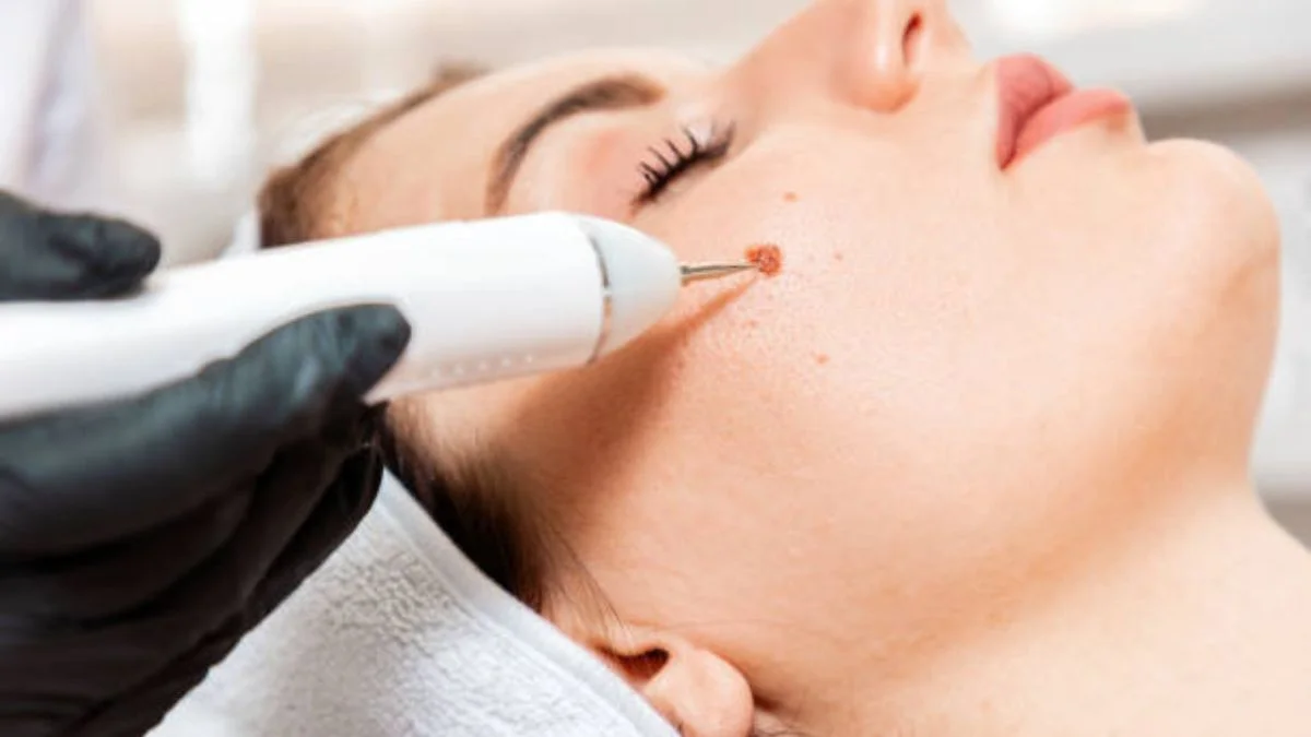
Skin Lesion Removal in Riyadh relies heavily on the integration of clinical expertise and accurate laboratory analysis, a process known as dermatopathology. While many lesions can be identified visually, the microscopic examination of removed tissue is often the final, definitive step in ensuring a patient's long-term health. Dermatopathology is the intersection of dermatology and pathology, where specialized physicians examine skin samples to provide precise diagnoses. This diagnostic layer is critical because it moves beyond surface-level observation, allowing clinicians to understand the cellular architecture of a lesion. For patients, this means that every removal—whether for cosmetic improvement or clinical necessity—is underpinned by a commitment to diagnostic rigor, ensuring that any underlying pathology is identified and treated with the necessary precision.
The primary purpose of dermatopathology is to provide a definitive diagnosis that guides further management. When a lesion is removed, it is standard practice to submit the excised tissue to a laboratory for histological examination. Under a microscope, a pathologist assesses the tissue at a cellular level, looking for specific architectural features, cellular changes, and the margins of the excision. This is particularly important for pigmented lesions or any growth that has exhibited rapid change in size, shape, or color. By analyzing the margins, pathologists can determine if a lesion was completely removed, which is vital for preventing recurrence. For the patient, this process provides peace of mind, confirming that the treatment plan was based on verified data rather than just clinical suspicion alone.
The journey of a tissue sample begins immediately after the excision. Once the skin lesion is removed, it is carefully preserved in a fixative solution and transported to a laboratory. There, it undergoes a meticulous process of tissue processing, embedding, and sectioning. The tissue is sliced into ultra-thin segments, mounted on glass slides, and stained to highlight specific cellular components. This allows the dermatopathologist to observe the skin’s layers—the epidermis, dermis, and sometimes subcutaneous fat—in extreme detail. This systematic approach is designed to catch even the most subtle abnormalities that are invisible to the naked eye. In Riyadh, the implementation of these high-standard laboratory workflows ensures that every patient benefits from a robust and standardized diagnostic path, regardless of the simplicity or complexity of the lesion.
Clinical diagnosis is a vital skill, but it is not infallible. Some benign lesions can mimic malignant ones, and conversely, early-stage cancers can sometimes present as innocuous bumps or patches. This is where the diagnostic partnership between the clinician and the pathologist becomes invaluable. If a clinician is uncertain about the nature of a lesion, they may opt for a diagnostic biopsy—a small sample taken to confirm the diagnosis—before performing a full excision. This strategy minimizes unnecessary surgical trauma and ensures that if a more extensive procedure is required, it is planned with the benefit of an accurate pathology report. This methodical approach is the hallmark of modern dermatological care, focusing on minimizing patient risk while maximizing the utility of every diagnostic intervention.
The effectiveness of this process is heavily reliant on communication. When a lesion is submitted for testing, the clinician provides the pathologist with pertinent clinical history, such as the location of the lesion, its duration, and any physical characteristics observed during the examination. This context helps the pathologist tailor their assessment, focusing on the most relevant features. Once the report is generated, the findings are shared with the clinician, who then discusses the results with the patient. If the diagnosis reveals an unexpected condition, this communication loop allows for prompt adjustments to the treatment plan. This integrated care model ensures that the patient is supported throughout the entire duration of their care, from the initial concern to the final pathological verification.
In Riyadh’s rapidly evolving healthcare environment, adhering to standardized protocols for tissue handling and reporting is non-negotiable. These standards ensure that patients receive consistent, high-quality care that meets international benchmarks. By prioritizing the role of dermatopathology, the medical community maintains a high level of vigilance, ensuring that skin lesions are not only removed effectively but also analyzed thoroughly. This commitment to detail is what defines a professional and secure approach to dermatological services. As medical technologies continue to advance, the synergy between clinical observation and pathological analysis will remain the cornerstone of effective skin lesion removal, providing patients with the confidence that their health is being managed through evidence-based, thorough, and highly accurate practices.